Research / Cardiovascular Flow
Cardiovascular Flow
Investigating blood transport, vortex dynamics, and cardiovascular device interactions using clinical imaging, experimental flow measurements, and computational modeling.
Investigating blood transport, vortex dynamics, and cardiovascular device interactions using clinical imaging, experimental flow measurements, and computational modeling.
Clinical ultrasound and patient-specific experimental models reveal how disease and mechanical circulatory support influence ventricular blood transport and aortic flow.
Rossini, L., et al. (2016). “A clinical method for mapping and quantifying blood stasis in the left ventricle.” Journal of Biomechanics, 49(11), 2152–2161.
Rossini, L., et al. (2021). “Non-invasive mapping of intraventricular flow patterns in patients treated with left ventricular assist devices.” ASAIO Journal, 67(1), 74–83.
Patient-specific flow fields are experimentally evaluated through a collaboration with Houston Methodist Hospital and the University of Texas, Houston Medical Center.
Our published 2-D PIV studies quantify how vortex structure and strength change with LVAD support. Current volumetric measurements extend this work to three-dimensional vortex-ring formation and evolution.
Wong, K., G. Samaroo, I. Ling, W. Dembitsky, R. Adamson, J. C. del Alamo, and K. May-Newman. “Intraventricular flow patterns and stasis in the LVAD-assisted heart.” Journal of Biomechanics 47(6):1485–1494, 2014. View paper ↗
Ortiz, S., V. Vu, R. Montes, and K. May-Newman. “Left Ventricular Flow Dynamics with the HeartMate3 Left Ventricular Assist Device: Effect of Inflow Cannula Position and Speed Modulation.” ASAIO Journal 67(12):1301–1311, 2021.
Vu, V., L. Rossini, J. C. del Alamo, W. Dembitsky, R. A. Gray, and K. May-Newman. “Benchtop models of patient-specific intraventricular flow during heart failure and LVAD support.” ASME Journal of Biomechanical Engineering 145(11):111010, 2023.
We use experimental flow fields to characterize cardiovascular transport and evaluate computational predictions, building on collaborative VVUQ research with the FDA and Barcelona Supercomputing Center.

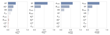
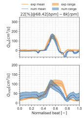
Santiago, A., et al. (2022). “Design and execution of a Verification, Validation, and Uncertainty Quantification plan for a numerical model of left ventricular flow after LVAD implantation.” PLOS Computational Biology, 18(6), e1010141. View publication ↗
The PIV and CFD panels illustrate complementary methods; their displayed frames are not a validated point-by-point matched comparison. Final matched imagery and CFD video processing remain to be completed.